Silk from the tasar silkworm used as a scaffold for heart tissue
 Since almost all of the body’s own regeneration mechanisms in the heart have become deactivated, a heart attack or other heart damage is serious for patients since the dead cardiac cells are irretrievably lost and scar tissue grows in place of the damaged muscle cells. Max Planck Institute for Heart and Lung Research in Bad Nauheim researchers are on their way to find a solution for this problem.
They are pursuing development of an artificial cardiac tissue that could be grown in a laboratory and used as patches for damaged cardiac muscles. There have been many experiments in which different materials have been tested as a potential scaffold substance for the loading of cardiac muscle cells, however, the main problem thus far was the reconstruction of a three-dimensional structure that would be suitable for long term use along this hard working organ.
“Whether natural or artificial in origin, all of the tested fibers had serious disadvantages”, said Felix Engel, Research Group Leader at the Max Planck Institute for Heart and Lung Research in Bad Nauheim. “They were either too brittle, were attacked by the immune system or did not enable the heart muscle cells to adhere correctly to the fibers.”
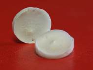
However, the scientists have now found a possible solution at the University of Kharagpur, India, where researchers managed to produce coin-sized disks from the cocoon of the tasar silkworm (Antheraea mylitta). According to Chinmoy Patra, an Indian scientist who now works in Engel’s laboratory, the fiber produced by the tasar silkworm displays several advantages over the other substances tested.
“The surface has protein structures that facilitate the adhesion of heart muscle cells. It’s also coarser than other silk fibers”, said Engel. “The communication between the cells was intact and they beat synchronously over a period of 20 days, just like real heart muscle.”
By using the silk produced by a tropical silkworm, the researchers were able to load cardiac muscle cells onto a three-dimensional scaffold. Although the results look promising, the researchers claim that the clinical application of the fiber on humans won’t be available in the near future.
“Unlike in our study, which we carried out using rat cells, the problem of obtaining sufficient human cardiac cells as starting material has not yet been solved”, said Engel.
For more information, read the paper published in the journal Biomaterials: “Silk protein fibroin from Antheraea mylitta for cardiac tissue engineering”.


Application of BIOMIMICRY.
Dr.A.Jagadeesh Nellore (AP), India
E-mail: anumakonda.jagadeesh@gmail.com