The interactive 3D Virtual Autopsy Table (Inside Explorer)
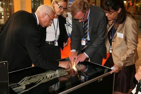
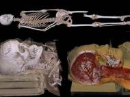 Swedish researchers have developed an interactive touchscreen 3D autopsy table that allows pathologists to examine virtual representations of real bodies in minute detail and from different viewing angles. The Virtual Autopsy Table has been developed by Norrköping Visualization Center in cooperation with Center for Medical Image Science and Visualization using science and technology which is already used for other purposes.
The victim’s body is placed on an examination table under a computed tomography (CT) scanner and/or magnetic resonance imaging (MRI) machine and processed using software developed by the researchers. A CT scan takes only 20 seconds and displays the bones, gases and any foreign objects in the body. A specially-developed technique known as quantative synthetic MRI allows for scanning of dead bodies and provides data on soft tissue. The software converts the layer by layer data sets provided by the scans and builds a 3D virtual visualization of the victim’s body.
The visualization allows an examiner to look at a body in microscopic detail. Going inside the body is simply a matter of removing the virtual skin and muscle layers to reveal the skeleton and organs. The examiner can zoom in and out, view cross-sections using a virtual scalpel and control the level of layer transparency with relative ease.
Whereas an actual invasive autopsy can take some time to complete, the cause of death using a virtual autopsy could be established in as little as 15 minutes. Examining injuries like bone fractures can be very complicated and current photographic evidence-gathering is limited. However, using the visualization techniques can offer a unique viewing opportunity and accurately show what an injury looks like.
Photographic evidence presented in criminal prosecutions can be difficult to explain to those unfamiliar with pathology and can often make for shocking viewing. Showing the evidence as a 3D representation of the victim makes explanations more relevant and easier to understand. Researchers claim to have proven that the virtual autopsies provide more information than their real-world counterparts. The table can be used for living patients as well, so it can give a better picture of injury or illness progress to specialists.
The Project Manager Thomas Rydell said: “We are currently using an LCD-based diffuse illumination multitouch table, it can handle multiple tracking points and fiducials. The volume renderer developed at Linköping University allows us to do the rendering at interactive frame rates with very low latency on a regular gaming card, in this case a NVIDIA GTX 295, the fastest gaming card on the market. The rendering is done in full HD resolution.”
The project team currently has only one working mobile demonstration table but is looking to install tables in a number of selected public institutions from next year which will no doubt be of great interest to the students of anatomy and, of course, technology lovers. The working demonstration model currently travels the globe to be shown at various technology and healthcare conferences and events.





MEMPHIS TENNESSEE US OF A NEEDS ONE. SELL THEM A TABLE.
from where can I purchase this table please let me know.
author
The new name of Virtual Autopsy Table is Inside Explorer, and it is suitable for exhibitions at museums or science centers. They also made Sectra Visualization Table which is a version more suitable for use in medical purposes.